Tendons And Ligaments In Foot And Leg : Calcaneus bone anatomy, function, calcaneus pain ... - All of these nerves extend as branches of nerves in the leg that pass through the ankle and into the foot.
bymamadeerdoff-
0
Tendons And Ligaments In Foot And Leg : Calcaneus bone anatomy, function, calcaneus pain ... - All of these nerves extend as branches of nerves in the leg that pass through the ankle and into the foot.. The sural nerve branches from the tibial and common fibular nerves and is responsible for feeling on the outside of the foot and the small toe. We have information on sports injuries with treatment, rehabilitation, exercises, strapping & taping and more. All of these nerves extend as branches of nerves in the leg that pass through the ankle and into the foot. Bands of connective tissue called retinacula (singular: The plantar aspect of the foot contains the tough fibrous plantar aponeurosis covering muscles and tendons arranged in 4 layers, numbered from 1 superficial to 4 deep:
The sural nerve branches from the tibial and common fibular nerves and is responsible for feeling on the outside of the foot and the small toe. A number of tendons pass through the ankle region. The lateral ligaments, the deltoid ligament on the medial side, and the ligaments of the tibiofibular syndesmosis that join the distal epiphyses of the bones of the leg (tibia and fibula). The tendons in the foot are highly complex and intricate. Tendons are the main collagenous structures in the dorsum.
Leg Anatomy Muscles And Tendons How To Fix Achilles ... from i.pinimg.com Therefore, the healing process for a broken tendon is long and painful. The plantar aspect of the foot contains the tough fibrous plantar aponeurosis covering muscles and tendons arranged in 4 layers, numbered from 1 superficial to 4 deep: We have information on sports injuries with treatment, rehabilitation, exercises, strapping & taping and more. Jul 25, 2017 · in humans, the foot is one of the most complex structures in the body. Bands of connective tissue called retinacula (singular: Retinacula, tendons and their synovial sheaths, vessels, and nerves. The tendons connect anterior/dorsiflexor compartment muscles of the leg to the foot bones. Mar 23, 2010 · the ligaments around the ankle can be divided, depending on their anatomic position, into three groups:
Bands of connective tissue called retinacula (singular:
The lateral ligaments, the deltoid ligament on the medial side, and the ligaments of the tibiofibular syndesmosis that join the distal epiphyses of the bones of the leg (tibia and fibula). Mnemonics that can be used to remember the anatomy of the ankle tendons from anterior to posterior as they pass posterior to the medial malleolus of the tibia under the flexor retinaculum in the tarsal tunnel include: The sural nerve branches from the tibial and common fibular nerves and is responsible for feeling on the outside of the foot and the small toe. We have information on sports injuries with treatment, rehabilitation, exercises, strapping & taping and more. A number of tendons pass through the ankle region. Therefore, the healing process for a broken tendon is long and painful. Bands of connective tissue called retinacula (singular: Mar 23, 2010 · the ligaments around the ankle can be divided, depending on their anatomic position, into three groups: Examples that help finger movement include: The tendons in the foot are highly complex and intricate. The plantar aspect of the foot contains the tough fibrous plantar aponeurosis covering muscles and tendons arranged in 4 layers, numbered from 1 superficial to 4 deep: Jul 03, 2018 · the nerves of the foot help move the body and keep balance both while it's moving and at rest. Jul 25, 2017 · in humans, the foot is one of the most complex structures in the body.
The lateral ligaments, the deltoid ligament on the medial side, and the ligaments of the tibiofibular syndesmosis that join the distal epiphyses of the bones of the leg (tibia and fibula). The plantar aspect of the foot contains the tough fibrous plantar aponeurosis covering muscles and tendons arranged in 4 layers, numbered from 1 superficial to 4 deep: Mar 18, 2020 · ligaments hold your bones together, and although they can't be strengthened directly, targeting the muscles around them will strengthen your tendons and improve foot stability. Jul 25, 2017 · in humans, the foot is one of the most complex structures in the body. Mar 23, 2010 · the ligaments around the ankle can be divided, depending on their anatomic position, into three groups:
What is a Retinaculum? from corewalking.com Many tendons in your hands and feet attach to hand and foot bones, and help you move your fingers and toes. Welcome to the virtual sports injury clinic. Tendons are the main collagenous structures in the dorsum. The plantar aspect of the foot contains the tough fibrous plantar aponeurosis covering muscles and tendons arranged in 4 layers, numbered from 1 superficial to 4 deep: A number of tendons pass through the ankle region. We have information on sports injuries with treatment, rehabilitation, exercises, strapping & taping and more. The tendons in the foot are highly complex and intricate. Examples that help finger movement include:
The tendons connect anterior/dorsiflexor compartment muscles of the leg to the foot bones.
Among these important structures is the achilles tendon — attaching your calf muscles to your heel bone. A number of tendons pass through the ankle region. The lateral ligaments, the deltoid ligament on the medial side, and the ligaments of the tibiofibular syndesmosis that join the distal epiphyses of the bones of the leg (tibia and fibula). Welcome to the virtual sports injury clinic. Mnemonics that can be used to remember the anatomy of the ankle tendons from anterior to posterior as they pass posterior to the medial malleolus of the tibia under the flexor retinaculum in the tarsal tunnel include: Therefore, the healing process for a broken tendon is long and painful. The plantar aspect of the foot contains the tough fibrous plantar aponeurosis covering muscles and tendons arranged in 4 layers, numbered from 1 superficial to 4 deep: Most people who do not receive medical attention within the first 48 hours of the injury will suffer from severe swelling, pain, and a burning sensation where the injury occurred. Bands of connective tissue called retinacula (singular: Mar 18, 2020 · ligaments hold your bones together, and although they can't be strengthened directly, targeting the muscles around them will strengthen your tendons and improve foot stability. The tendons in the foot are highly complex and intricate. The sural nerve branches from the tibial and common fibular nerves and is responsible for feeling on the outside of the foot and the small toe. Retinacula, tendons and their synovial sheaths, vessels, and nerves.
The sural nerve branches from the tibial and common fibular nerves and is responsible for feeling on the outside of the foot and the small toe. Most people who do not receive medical attention within the first 48 hours of the injury will suffer from severe swelling, pain, and a burning sensation where the injury occurred. The tendons connect anterior/dorsiflexor compartment muscles of the leg to the foot bones. Welcome to the virtual sports injury clinic. All of these nerves extend as branches of nerves in the leg that pass through the ankle and into the foot.
Muscles that lift the Arches of the Feet from corewalking.com Tendons are the main collagenous structures in the dorsum. Bands of connective tissue called retinacula (singular: We have information on sports injuries with treatment, rehabilitation, exercises, strapping & taping and more. Mnemonics that can be used to remember the anatomy of the ankle tendons from anterior to posterior as they pass posterior to the medial malleolus of the tibia under the flexor retinaculum in the tarsal tunnel include: The tendons connect anterior/dorsiflexor compartment muscles of the leg to the foot bones. Mar 18, 2020 · ligaments hold your bones together, and although they can't be strengthened directly, targeting the muscles around them will strengthen your tendons and improve foot stability. Many tendons in your hands and feet attach to hand and foot bones, and help you move your fingers and toes. All of these nerves extend as branches of nerves in the leg that pass through the ankle and into the foot.
All of these nerves extend as branches of nerves in the leg that pass through the ankle and into the foot.
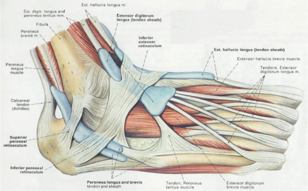
Mnemonics that can be used to remember the anatomy of the ankle tendons from anterior to posterior as they pass posterior to the medial malleolus of the tibia under the flexor retinaculum in the tarsal tunnel include: The plantar aspect of the foot contains the tough fibrous plantar aponeurosis covering muscles and tendons arranged in 4 layers, numbered from 1 superficial to 4 deep: Mar 18, 2020 · ligaments hold your bones together, and although they can't be strengthened directly, targeting the muscles around them will strengthen your tendons and improve foot stability. We have information on sports injuries with treatment, rehabilitation, exercises, strapping & taping and more. The lateral ligaments, the deltoid ligament on the medial side, and the ligaments of the tibiofibular syndesmosis that join the distal epiphyses of the bones of the leg (tibia and fibula). The sural nerve branches from the tibial and common fibular nerves and is responsible for feeling on the outside of the foot and the small toe. Retinaculum) allow the tendons to exert force across the angle between the leg and foot without lifting away from the angle, a process called bowstringing. Most people who do not receive medical attention within the first 48 hours of the injury will suffer from severe swelling, pain, and a burning sensation where the injury occurred. Retinacula, tendons and their synovial sheaths, vessels, and nerves. Bands of connective tissue called retinacula (singular: The tendons in the foot are highly complex and intricate. Jul 03, 2018 · the nerves of the foot help move the body and keep balance both while it's moving and at rest. The tendons connect anterior/dorsiflexor compartment muscles of the leg to the foot bones.